A sudden dashboard impact, a sports collision, or a hard fall can push the shin backward and injure the posterior cruciate ligament. When this strong band tears, the knee may feel unstable, swollen, or strangely loose while walking downstairs. Some people notice little pain at first. That can delay proper assessment.
PCL reconstruction surgery rebuilds a severely damaged posterior cruciate ligament with a tendon graft. The graft may come from the patient or a donor. Orthopaedic knee specialist Robert F. LaPrade, MD, describes the PCL as “the primary restraint to posterior translation of the tibia.” His point matters because the ligament controls more than simple forward movement. It also supports coordinated knee motion during turning, squatting, and deceleration.
Not every PCL tear needs surgery. A partial tear, stable knee, or improving function may respond to bracing and structured physical therapy. Surgery becomes more likely when instability persists, the ligament is completely torn, or other ligaments and cartilage are injured. Examination, stress radiographs, and MRI help clarify the damage. Still, scans do not replace a careful physical assessment.
PCL reconstruction surgery is not a guaranteed return to sport. Recovery often involves weeks of protected movement, gradual strengthening, and patient rehabilitation. Early bending can be limited. Progress can feel slow. That is normal, but not always harmless. Each case differs, and treatment decisions should reflect symptoms, activity goals, age, and associated injuries. A thoughtful consultation can reveal whether reconstruction is truly necessary—or whether a less invasive path deserves more time.
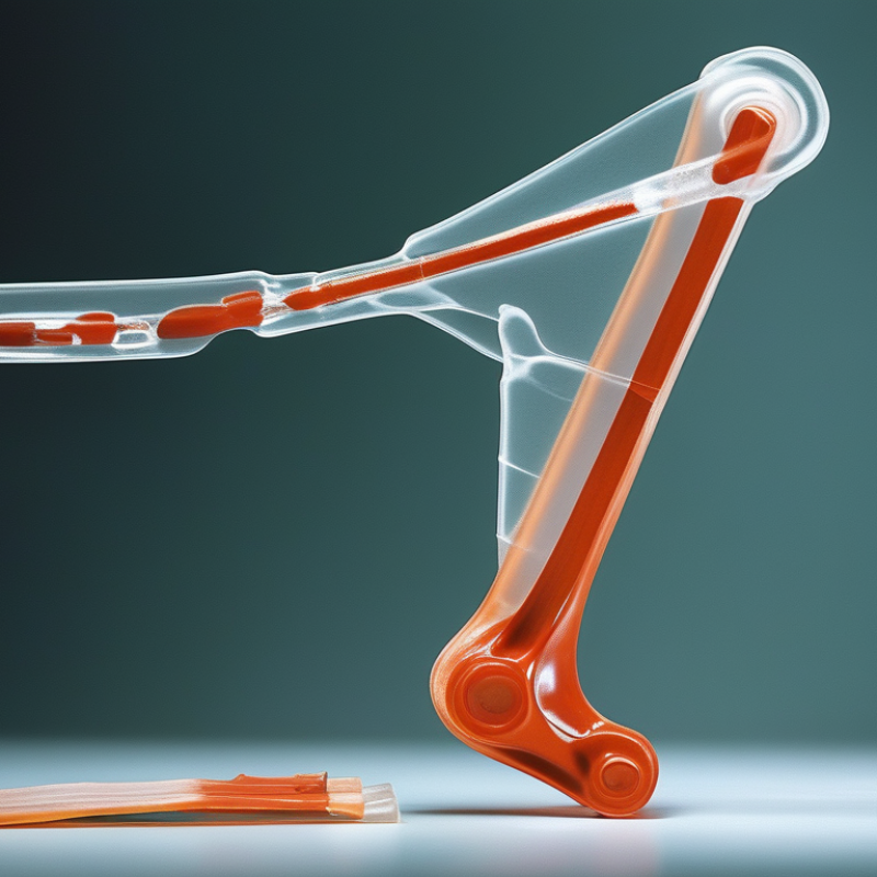
The posterior cruciate ligament, or PCL, sits deep inside the knee. It connects the thighbone to the shinbone. Its main job is to stop the shinbone from moving too far backward. It also helps control knee rotation during walking, running, and turning. Think of it as a strong internal strap that guides movement when the knee bends. The knee still moves.
A PCL injury often follows a forceful blow to the front of a bent knee. It can also occur after a fall, sports collision, or awkward landing. Some people notice swelling, stiffness, or pain behind the knee. Others mainly feel that the knee shifts on stairs or uneven ground. Pain alone does not prove a PCL tear. That distinction matters, and it is easy to miss. A clinician usually reviews the injury, checks knee stability, and may request magnetic resonance imaging or X-rays.
Not every PCL injury needs reconstruction surgery. Mild or isolated tears may improve with guided rehabilitation, strength work, and activity changes. Surgery may be considered when instability continues, rehabilitation fails, or other knee ligaments are also damaged. People with demanding work or sports goals may need a more detailed discussion. Recovery is not identical for everyone. Even accurate imaging cannot replace a physical examination, and treatment decisions should account for symptoms, movement tests, health conditions, and personal goals.
A posterior cruciate ligament (PCL) injury often follows a forceful blow to the front of the knee. A dashboard impact during a crash is a classic example. A hard fall onto a bent knee can also damage the ligament. Contact sports, sudden hyperextension, and twisting may cause less obvious injuries. Some people remember a sharp pop. Others notice swelling, stiffness, or a knee that feels unstable days later. That detail matters.
Diagnosis begins with the injury history and a careful physical examination. A clinician asks how the knee was struck, when swelling appeared, and whether walking feels unsafe. During examination, the knee is checked for looseness and abnormal movement. The posterior drawer test measures backward movement of the shinbone. A visible “sag” may also suggest PCL damage. Pain and swelling can make these tests difficult. No single test is perfect.
X-rays can identify fractures or changes in bone position, but they cannot show the PCL clearly. Magnetic resonance imaging, often called MRI, can reveal a partial or complete tear. It may also show damage to cartilage, the meniscus, or other ligaments. In complex cases, clinicians compare both knees and review earlier images. Even experienced examiners can miss a combined injury. A second assessment may be sensible when instability continues, especially before considering PCL reconstruction surgery.
A posterior cruciate ligament injury is commonly graded by the amount of posterior tibial translation during clinical examination. Grade I indicates 1–5 mm, Grade II indicates 6–10 mm, and Grade III indicates more than 10 mm.
Diagnosis usually combines a posterior drawer test with imaging. Stress radiographs can quantify instability, while MRI helps evaluate the PCL and associated injuries. The grade alone does not determine whether reconstruction surgery is needed; symptoms, instability, activity level, and additional knee damage are also considered.
PCL reconstruction surgery is considered when the posterior cruciate ligament cannot reliably stabilize the knee. Not every tear needs reconstruction. Published epidemiology reviews estimate PCL injuries represent about 1–3% of knee ligament injuries, although registry definitions vary (Journal of Orthopaedic Surgery and Research, 2019). The American Academy of Orthopaedic Surgeons recommends assessing symptoms, knee looseness, associated injuries, and activity demands before choosing surgery.
Surgery is more likely after a complete tear causes repeated buckling, difficulty walking downhill, or pain during twisting. It may also be recommended when the PCL tear occurs with ACL, collateral ligament, meniscus, or cartilage damage. A displaced PCL avulsion fracture may require fixation instead of reconstruction. Persistent instability after structured rehabilitation is another important trigger. The decision is not perfectly neat. MRI alone cannot measure functional instability.
Tips: Ask whether your knee has measurable posterior laxity. Request an explanation of stress radiographs and rehabilitation timelines. Discuss non-surgical care when the injury is isolated and symptoms are manageable. Early treatment matters most in multiligament injuries, especially when circulation or nerve function is affected.
A sports-medicine specialist should compare both knees during examination. Strength, gait, swelling, and movement patterns all matter. Current reviews in Knee Surgery, Sports Traumatology, Arthroscopy emphasize individualized treatment, because patient goals and injury combinations strongly influence outcomes. Recovery commonly requires several months, and returning too quickly can undermine stability. Pain may improve before control does. That difference deserves attention.
PCL reconstruction surgery repairs a torn posterior cruciate ligament in the knee. It may be recommended when the tear causes ongoing instability, pain, or difficulty walking. During the operation, the surgeon usually uses a tendon graft to replace the damaged ligament. The graft may come from the patient’s own body or a screened donor.
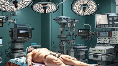
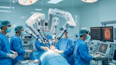
The patient receives anesthesia and lies in a carefully supported position. Small openings allow an arthroscope and surgical instruments to enter the knee. A camera displays the joint on a monitor, helping the surgeon inspect cartilage, menisci, and other ligaments. Damaged tissue may be treated during the same procedure. The surgeon then prepares narrow bone tunnels in the thighbone and shinbone. The graft passes through these tunnels and is secured with fixation devices. Precise tension matters. Too loose can leave the knee unstable; too tight may restrict movement.
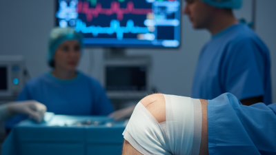
After surgery, the knee is placed in a brace, and walking may require crutches. Physical therapy usually begins with gentle motion and swelling control. Strength and balance exercises progress gradually over several months. Recovery is not perfectly linear. A good day can be followed by stiffness or swelling, which can feel discouraging. That does not always mean the reconstruction has failed, but new instability, fever, severe pain, or calf swelling needs prompt medical review. The surgical plan and rehabilitation schedule should match the person’s injury, health, and activity goals.
| Data Dimension | Key Information | Typical Details | Clinical Considerations |
|---|---|---|---|
| Definition | Posterior cruciate ligament (PCL) reconstruction is an operation that replaces a torn PCL with a tendon graft to help restore knee stability. | The graft may be obtained from the patient or from screened donor tissue. | It is different from PCL repair, which attempts to preserve and reattach the original ligament. |
| Function of the PCL | The PCL connects the thighbone to the shinbone and helps prevent the shinbone from moving too far backward relative to the thighbone. | It contributes to stability during walking, turning, squatting, and activities that load the bent knee. | PCL injuries can occur alone or together with damage to other knee ligaments, cartilage, or the meniscus. |
| Common Injury Mechanisms | A force directed to the front of the shin while the knee is bent is a classic mechanism of PCL injury. | Other causes include a fall onto a bent knee, sports-related twisting, or knee dislocation. | High-energy injuries require careful assessment for fractures, vascular injury, nerve injury, and additional ligament damage. |
| Who May Need Surgery? | Surgery may be considered for persistent instability, severe or complete tears, combined ligament injuries, or symptoms that do not improve with appropriate nonsurgical care. | Treatment decisions depend on activity goals, knee stability, injury pattern, symptoms, and overall health. | Not every isolated PCL tear requires reconstruction; some improve with bracing, rehabilitation, and activity modification. |
| Preoperative Assessment | The clinician reviews the injury history, examines knee stability and alignment, and evaluates movement and strength. | Imaging may include X-rays and magnetic resonance imaging (MRI). | Swelling, reduced range of motion, untreated infection, or other medical issues may affect surgical timing. |
| Anesthesia and Positioning | The procedure is usually performed under regional or general anesthesia, depending on the patient and anesthesiology plan. | The patient is positioned so the surgical team can examine and access the knee safely. | The exact anesthesia plan and surgical approach are individualized. |
| Arthroscopic Examination | Small incisions allow a camera and instruments to be inserted into the knee joint. | The surgeon assesses the PCL, other ligaments, menisci, cartilage, and joint surfaces. | Additional injuries may be treated during the same operation when appropriate. |
| Graft Selection | A graft is prepared to replace the damaged PCL. | Possible sources include the patient’s own tendon or carefully screened donor tissue. | Choice depends on surgeon assessment, patient factors, associated injuries, and informed consent. |
| Tunnel Preparation and Graft Placement | Bone tunnels are created in the thighbone and shinbone at or near the PCL’s anatomical attachment sites. | The graft is passed through the tunnels and positioned to reproduce the ligament’s stabilizing function. | Tunnel placement and graft tension are important for stability and knee motion. |
| Fixation and Closure | The graft is secured in place with fixation devices selected by the surgical team. | Incisions are closed and covered with dressings; a brace may be applied. | The operation may be more extensive when multiple ligaments require treatment. |
| Early Recovery | Initial care focuses on pain control, swelling reduction, wound care, protected weight-bearing, and restoring safe knee motion. | Crutches and a hinged knee brace are commonly used during the early phase. | Weight-bearing and range-of-motion limits vary according to the reconstruction and any additional procedures. |
| Rehabilitation | Physical therapy gradually develops motion, quadriceps strength, balance, coordination, and functional control. | Progression is based on healing, examination findings, strength, and functional milestones rather than time alone. | Returning too quickly to pivoting, jumping, or contact activities can increase the risk of reinjury. |
| Potential Risks | Possible complications include infection, bleeding, blood clots, stiffness, persistent pain, numbness, reduced motion, graft failure, or continued instability. | Risk varies with the injury, surgical complexity, medical history, and rehabilitation. | Urgent medical review is needed for severe calf swelling or pain, chest pain, breathing difficulty, fever, wound drainage, or sudden worsening symptoms. |
Recovery after PCL reconstruction is gradual and closely supervised. During the first days, most patients use crutches and a hinged brace. The knee may feel swollen, stiff, and surprisingly weak. Elevating the leg and following prescribed pain-control instructions can help. Your surgeon decides when weight-bearing is safe.
Physical therapy usually begins early, often with gentle motion and quadriceps activation. Heel slides, ankle movements, and supported leg raises may seem simple. They are not always easy. I have found that small improvements can feel invisible from one day to the next. That frustration is normal, but forcing movement can delay healing. Follow-up visits help the medical team adjust exercises, brace settings, and activity limits.
Weeks later, therapy may include controlled strengthening, balance work, and stationary cycling. Running, jumping, and sports require stronger muscles and reliable knee control, not just a certain date on the calendar. Recovery is rarely perfectly linear. A good week can be followed by stiffness or swelling. Contact your clinical team about increasing pain, wound drainage, fever, calf tenderness, or sudden shortness of breath. These symptoms need prompt medical assessment. Your rehabilitation plan should reflect the graft, associated injuries, examination findings, and daily progress.