>

5 Best Hip Arthroscopy Procedures for Global Buyers

Choosing among the five best hip arthroscopy procedures requires more than comparing prices, recovery times, or hospital brochures. Global buyers need evidence, surgeon experience, imaging quality, implant standards, and clear aftercare plans. Hip arthroscopy procedures may address labral tears, femoroacetabular impingement, cartilage damage, loose bodies, or selected ligament problems. Each option fits a different clinical picture.

Dr. Marc J. Philippon, a widely recognized hip-preservation specialist, has described the central goal as “restoring the hip to its normal anatomy.” That principle matters. A technically impressive operation may still be unsuitable for a patient with advanced arthritis, severe bone loss, or unrealistic expectations. Details matter.

This guide examines five commonly discussed procedures through a practical, international lens. It considers indications, surgical goals, expected rehabilitation, equipment requirements, and questions buyers should ask providers. Patients may need crutches, supervised physiotherapy, and several months before demanding activities feel comfortable. Outcomes also vary between individuals.

A ranked list can oversimplify. Surgeon judgment remains essential.

Buyers should verify board certification, hospital accreditation, published outcomes, infection-control procedures, and communication support before choosing treatment abroad. They should also request itemized costs, including imaging, anesthesia, rehabilitation, travel, and possible follow-up care. No procedure guarantees pain-free movement. Still, careful selection can reduce avoidable uncertainty and support a safer, more informed decision.

5 Best Hip Arthroscopy Procedures for Global Buyers

FAI Correction: 80–90% Return to Sport (BJSM Reviews)

5 Best Hip Arthroscopy Procedures for Global Buyers

FAI Correction: 80–90% Return to Sport (BJSM Reviews)

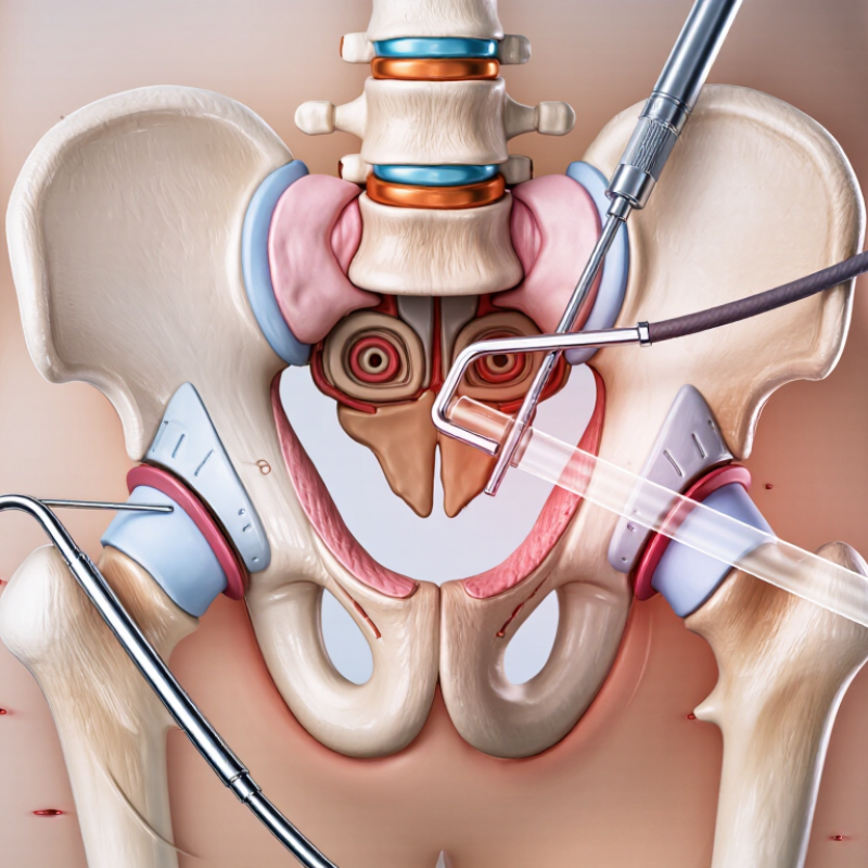
Femoroacetabular impingement, or FAI, occurs when abnormal bone shapes limit hip movement. Hip arthroscopy can correct these shapes through small incisions. Surgeons may trim a cam lesion, reshape a pincer lesion, or address both. They also inspect the labrum and cartilage during the procedure.

BJSM reviews report return-to-sport rates of approximately 80–90% after appropriate FAI treatment. This figure is encouraging, but it is not a promise. Results depend on cartilage health, symptom duration, athletic demands, surgical technique, and rehabilitation quality. A professional footballer and a recreational runner may follow different recovery plans.

The details matter.

Before surgery, patients usually receive clinical testing and imaging. A specialist should explain whether pain comes mainly from FAI, a labral tear, or another condition. After surgery, early movement protects joint mobility, while progressive strength work rebuilds control. Running and pivoting often return later, after strength and movement symmetry improve.

Recovery can feel uneven. Some athletes regain confidence before their hip is ready for full competition. Others need more time than expected. That uncertainty deserves honest discussion. Buyers comparing international care should verify surgeon training, hospital standards, imaging review, rehabilitation access, and follow-up arrangements. A lower advertised price should never replace clear clinical evidence and individualized planning.

5 Best Hip Arthroscopy Procedures for Global Buyers

Reported return-to-sport ranges after commonly performed hip arthroscopy procedures

FAI correction is frequently associated with an approximately 80–90% return-to-sport rate in published reviews. Outcomes vary according to cartilage damage, rehabilitation quality, athletic level, and the specific procedure performed. The ranges shown are literature-based estimates rather than guarantees for individual patients.

Labral Repair: 80%+ Two-Year Improvement (Hip Preservation Studies)

Labral repair is a key hip preservation procedure for patients with a torn acetabular labrum. The labrum is a ring of cartilage that supports hip stability and joint movement. Hip preservation studies report more than 80% improvement at two years in selected patients. However, this figure usually reflects pain or function scores, not a guaranteed cure. Results depend on tear location, cartilage health, bone shape, activity level, and rehabilitation quality.

During repair, a surgeon reattaches the damaged tissue using small anchors. Some tears require reshaping of the hip socket or femur to reduce repeated impingement. Patients often use crutches briefly and begin guided exercises soon after surgery.

Full sporting recovery may take several months. My concern is simple: online success rates can hide patient selection. A healthy joint and an experienced surgeon matter greatly.

Tips: Ask for imaging copies and a written diagnosis before travelling. Confirm the surgeon’s training in hip preservation and arthroscopy. Request clear details about anesthesia, rehabilitation, follow-up, and possible revision surgery. A remote consultation can help, but it cannot replace a physical examination. Bring previous scans, medication records, and a realistic activity goal. Recovery rarely feels perfectly linear. Pain may improve before strength returns, and setbacks can occur.

Microfracture: Focal Cartilage Lesions Under 2 cm² (AAOS)

Microfracture is a focused hip arthroscopy procedure for small, contained cartilage lesions under 2 cm². It is commonly considered when the defect is localized and the surrounding joint remains reasonably healthy.

During arthroscopy, the surgeon gently prepares the damaged area and creates tiny openings in the underlying bone. Marrow elements then form a clot over the lesion. Over time, this may develop into fibrocartilage.

The 2 cm² threshold is useful, but it is not a magic line. Lesion depth, location, hip stability, bone shape, and patient activity also influence the decision. A surgeon should assess imaging alongside symptoms and physical findings. Pain from impingement may require correction during the same operation. Treating the cartilage alone can be insufficient.

Recovery demands patience. Early weight-bearing restrictions may protect the healing area, followed by controlled motion and progressive strengthening. Rehabilitation often takes several months. Outcomes are less predictable when arthritis, diffuse cartilage loss, or untreated structural problems are present. The evidence is still imperfect. Patients traveling internationally should verify the surgeon’s hip arthroscopy experience, facility standards, rehabilitation plan, and long-term follow-up access. Ask how lesion size was measured and what alternatives remain if microfracture does not provide lasting relief.

Loose-Body Removal: 90%+ Relief (Arthroscopy Case Series)

Loose-Body Removal: 90%+ Relief in an Arthroscopy Case Series

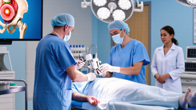
Loose bodies are small fragments of cartilage or bone moving inside the hip joint. They may cause catching, sharp pain, stiffness, or a sudden locking sensation. During hip arthroscopy, a surgeon makes small incisions and inserts a camera into the joint. Specialized instruments can then remove the fragment without opening the entire hip.

In one arthroscopy case series, more than 90% of patients reported meaningful relief after loose-body removal. This figure is encouraging, but it is not a guarantee. Results depend on the fragment’s size, joint damage, symptoms, and overall health. A careful examination and imaging review should come first.

Patients may describe pain when climbing stairs, turning in bed, or entering a car. These details help distinguish loose bodies from labral tears, arthritis, or muscle problems.

Recovery often includes crutches, controlled exercises, and gradual weight-bearing. Physical therapy may restore hip motion and strength. However, removing the loose body does not always repair existing cartilage damage. That limitation deserves honest discussion.

Some patients improve quickly, while others need several weeks before daily movement feels comfortable. Even a technically successful procedure can disappoint if the original diagnosis was incomplete. A qualified orthopedic specialist should explain expected benefits, possible complications, follow-up plans, and alternatives before treatment.

Synovectomy: 70–90% Short-Term Improvement (ISHA Evidence)

Synovectomy is a hip arthroscopy procedure that removes inflamed synovial tissue from the joint. Surgeons usually consider it when synovitis causes persistent pain, swelling, or restricted movement. Through small portals, a camera guides instruments inside the hip. Patients may notice reduced groin pain during walking, climbing stairs, or sitting.

ISHA evidence reports 70–90% short-term improvement in selected patients after synovectomy. The figure is encouraging, but it is not a promise. Results depend on the underlying condition, cartilage health, surgical technique, and rehabilitation. A patient with inflammatory synovitis may respond differently from someone with advanced arthritis. Clinical assessment, imaging, and medical history should guide the decision.

Recovery often includes protected weight-bearing, gentle range-of-motion exercises, and progressive strengthening. A physiotherapist may monitor hip flexion while the patient performs simple movements beside a treatment table. Complications are uncommon but can include infection, bleeding, nerve irritation, or continuing pain. I would also question any clinic that presents the 70–90% figure without defining “improvement” or its follow-up period. Short-term relief can fade when the original disease remains active. For international patients, verified surgeon credentials, transparent pricing, emergency planning, and realistic rehabilitation arrangements deserve careful attention.

5 Best Hip Arthroscopy Procedures for Global Buyers – Synovectomy: 70–90% Short-Term Improvement (ISHA Evidence)
Procedure Typical Indications What the Procedure Addresses Reported Short-Term Clinical Benefit Typical Rehabilitation Considerations Evidence and Buyer-Relevant Notes
Femoroacetabular Impingement (FAI) Osteoplasty Cam or pincer morphology with activity-related groin pain, restricted hip motion, and imaging-confirmed impingement. Reshapes excess bone at the femoral head–neck junction and/or acetabular rim to reduce abnormal contact. Most appropriately selected patients show meaningful improvement in pain and hip-function scores within 6–12 months. Protected weight-bearing is commonly used for approximately 2–6 weeks, depending on the extent of bone work and associated procedures. Strength and return-to-sport rehabilitation often continue for 3–6 months. Randomized trials have shown arthroscopic treatment can provide greater short-term improvement than structured physiotherapy for symptomatic FAI. Outcomes depend strongly on diagnosis, cartilage status, correction quality, and rehabilitation.
Acetabular Labral Repair Traumatic or degenerative labral tears with persistent symptoms, especially when the labral tissue is repairable and hip stability can be preserved. Uses sutures or anchors to restore the labrum’s seal and improve load distribution around the hip joint. Patient-reported pain and function commonly improve substantially during the first 6–12 months; repair is generally favored over removal when viable tissue can be preserved. Weight-bearing and hip-flexion limits are frequently used for the first 2–4 weeks. Full functional recovery may require 3–6 months, with longer timelines for high-impact sports. Clinical studies support labral preservation when technically feasible. Results are less predictable with advanced osteoarthritis, substantial cartilage loss, dysplasia, or untreated bony impingement.
Synovectomy Inflamed or hypertrophic synovium associated with inflammatory synovitis, proliferative synovial disorders, or selected cases of pigmented villonodular synovitis/tenosynovial giant-cell tumor. Removes diseased synovial tissue to reduce irritation, pain, swelling, and mechanical restriction. Approximately 70–90% short-term symptom or function improvement has been reported in selected cohorts; the range is not universal and recurrence risk varies by the underlying disease. Early motion is often encouraged, while weight-bearing restrictions depend on the disease, extent of excision, cartilage condition, and any additional procedure performed. International hip-arthroscopy literature describes meaningful short-term improvement in carefully selected patients. Long-term surveillance may be required for proliferative synovial disease because recurrence can occur.
Loose-Body Removal Intra-articular fragments caused by trauma, osteochondral injury, degenerative disease, synovial disorders, or developmental conditions. Removes mobile cartilage, bone, or calcified fragments that cause catching, locking, pain, or range-of-motion limitation. Mechanical symptoms may improve rapidly when the loose body is the principal pain generator and the joint has limited pre-existing cartilage damage. Recovery is often shorter than for procedures requiring bone reshaping or cartilage restoration, although weight-bearing may be modified when an osteochondral lesion is treated at the same time. The best results are associated with a clearly identified mechanical loose body and limited osteoarthritis. Removing fragments does not reverse established cartilage degeneration.
Acetabular Chondral Repair or Microfracture Small, contained full-thickness cartilage defects in patients without advanced diffuse osteoarthritis. Stimulates marrow-derived repair tissue through controlled perforations in the subchondral bone; may be combined with labral repair or FAI correction. Selected patients may experience meaningful pain and function improvement over 6–12 months, but the quality and durability of repair tissue are variable. Partial or restricted weight-bearing is commonly required for approximately 4–8 weeks, followed by gradual strengthening. Return to impact activity is usually delayed compared with isolated labral treatment. Microfracture is generally reserved for small, well-contained defects. Advanced arthritis, large lesions, poor bone quality, or untreated mechanical overload reduce the likelihood of durable benefit.
Clinical interpretation: Improvement percentages are approximate ranges reported in selected clinical studies and should not be treated as guaranteed results. Procedure selection should be based on symptoms, physical examination, radiographs, MRI or CT findings, cartilage status, hip morphology, patient age, activity goals, and surgeon assessment. “Short-term” generally refers to outcomes measured during the first 6–12 months after surgery.